- The gist of this article
In the article about Cholesterol and OPCs, Dr Jack Masquelier explained that oxidation of LDL-cholesterol is the root cause of most forms of cardiovascular disease and, eventually, of heart attacks. “We can prevent heart attacks,” said Masquelier, “thanks to antioxidative substances and OPCs are the type of substance that may prevent LDL from becoming oxidized. […] We learned that by inhibiting the oxidation of LDL, which is the first carrier of cholesterol, we can protect the vascular wall against a build-up of artherosclerotic plaque.” But, the cascade of events that may be set in motion when we fail to protect LDL-cholesterol against oxidation – with a fatal heart attack as its endpoint ‒ is highlighted in the following 10 points.
Table of contents
1. When cholesterol goes rancid.
When too many free radicals get in touch with too many unprotected LDLs, the free radicals will wreck the LDL and make its cholesterol turn rancid. Because cholesterol is a fatty substance, the radicals can turn it rancid just as they can make oils and butter “go bad.” In turn, rancid fats behave as free radicals in a chain reaction. That’s how rancidity spreads through butter, from the outside to the inside.
2. HDL leaves “emtpy handed.”
The rancid cholesterol is no longer recognized by the HDL as something it must pick up to take it to the liver. HDL departs from the site “emtpy handed” and leaves the oxidized cholesterol behind. In the meantime, the vascular wall has taken action.
3. Vascular wall in inflammatory frenzy.
Once struck by a free radical, the LDL and cholesterol cargo drives the vascular wall into an inflammatory frenzy. It considers the wrecked LDL an unwanted particle, an intruder that must be seriously dealt with. So, it calls for help. From the blood, the vascular wall attracts white blood cells, also known as leukocytes or monocytes. White blood cells act by moving through the vascular wall to reach a site of injury or to scavenge intruders or unknown particles. In this case, they come for oxidized cholesterol.
4. Vacuuming wrecked cholesterol.
Before moving through the wall, the white blood cells that have descended into the vascular wall must first mature into macrophages. When called to arms, these macrophages “vacuum” the wrecked LDL-cholesterol to clean up the area.
5. Foam cells.
Absorbing the excess of wrecked LDL-cholesterol, the macrophages become so laden with LDL’s fatty remainders that they become “foamy.” These fat-inflated foamcells get company from another white blood cell called a T-lymphocyte.
6. Coating the plaque.
This nightmare that began with free-radical-damaged LDL has now turned into an uncontrollable mess in which the wall’s muscle cells try to overgrow the mess by capping it with a coating of strong collagen fibers. Underneath it, the plaque may harden and go into “stenosis,” making the vascular wall inflexible.
7. The plaque breaks up.
The collagen cap may break or weaken under the influence of inflammatory agents. Collagen may also become brittle and break as a result of free radical attack. The plaque loses its cover.
8. Angina pectoris.
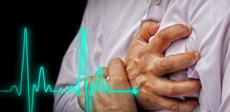
An open, uncapped plaque will then cause the release of clotting factors in the blood as well as in the plaque itself. When the blood clot stops the flow of blood in the heart muscle, a person feels pressure and tightness under the breastbone. When the clot persists, heart muscle tissue will remain without blood too long and it will acidify. This is experienced as “angina pectoris.”
9. Heart muscle without blood for too long.
In most cases, the acidification will take place in the left ventricle, which is the chamber of the heart where the muscle is thickest because it is this chamber that pumps the blood into the vascular system. The acidification will cause a stiffening of the red blood cells and in this condition they can no longer pass through the capillaries of the heart’s vascular system. Without a sufficient supply of red blood cells the muscle will be without oxygen too long.
10. Scars in heart muscle and heart attack.
Unless the acidification is undone, muscle cells may suffocate and die, releasing lysosomes, which are enzymes that destroy cells. What remains is a scar in the heart muscle. Too much scar tissue may stop the heart from functioning normally. In the worst case, when the muscle has become seriously damaged, the heart stops.
Make a fist and see for yourself
Most heart attacks find their origin in damage located in the muscle of the left ventricle. This is because the inside of the left ventricle’s muscle is particularly vulnerable when it comes to being without oxygen. With each beat of the heart, the ventricle’s muscle pushes against the blood. Because the blood in the chamber “pushes back,” the muscle, when it contracts, will squeeze itself bloodless for a brief moment. Then, it will refill itself, provided that the muscle’s vaculature is fully intact and operating normally. You can visualize this by making a fist with your hand and then squeeze hard. Open your hand and you’ll see how the blood will re-enter the muscles inside it.
Understandably, when, in the case of the heart, stiff red blood cells can’t get through the capillaries, the reperfusion of the heart’s muscle will stall and the heart will remain without blood for too long. This cascade of events begins with the failure to protect LDL-cholesterol against oxidation. So, as Dr. Jack Masquelier suggested, you may wish to consider keeping your LDL-cholesterol in good condition instead of trying to lower your cholesterol level and leave it unprotected against oxidation.


